Brain CT Scan v/s MRI Scan
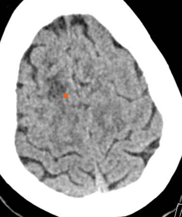
CT: The red dot refers to the dark area of the brain representing acute infarction.
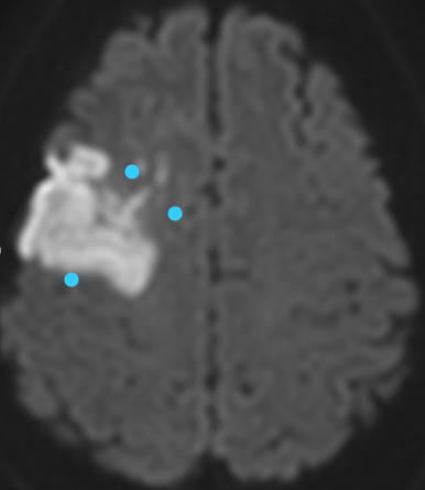
MRI: The blue dots refer to the bright area of the brain representing acute infarction.
Sometimes the terms CT Scan and MRI Scan are used interchangeably or referred to as simply a ‘scan’. However, there are several significant differences between the two and they are both often used in different situations.
I have tried to breakdown the technologies in simple terms.
What is a CT Scan?
Computerised Tomography or a CT Scan is a combination of several different X-ray images that are taken from different angles. A computer is used in the process to create an image by using these series of images that the X-ray generates.
A CT scan is preferred when time is of the essence. It is a quicker method and provides doctors with initial information to begin treatment, especially, in cases of medical emergencies such as trauma or a brain stroke.
What is an MRI Scan?
Magnetic Resonance Imaging or an MRI Scan uses powerful radio waves and magnetic fields to produce a very specific image. It can show minute details of the tissues of the body. It is considered the better technology when it comes to imaging.
An MRI scan is relied upon when the doctor needs to look at a detailed image of an organ, tissue or ligament such as cancer and infection. On the balance of probabilities depending on the case accurate characterisation of certain lesions is difficult to view with a CT scan, hence, doctors turn to an MRI scan. When it comes to brain MRI is usually far superior to CT.
CT Scan v/s MRI Scan
Both CT scan and MRI scan are advanced, modern healthcare technologies that have helped change the medical world for the better. It helps doctors and medical workers reach quicker and precise diagnosis which enables them to provide patients with quick, effective treatment. However both imaging still need to be interpreted in the light of clinical findings and laboratory reports. Otherwise the same images may have different differential diagnosis dependent on the background disease and the presentation of the signs and symptoms.
However, while both technologies are good at what they do, there are some significant differences between the two, like –
- CT Scans use radiation while MRI scans use radio waves and magnetic fields. Therefore MRI is on average counted as a safer method.
- MRI scans provide more detailed images while CT scans are not able to highlight the specific details of organs, tissues and ligaments.
- CT scans is non-invasive and, hence, painless; while MRI is also non-invasive, it may not be favoured for patients that have claustrophobia or trouble lying down for long.
- A CT scan is often preferred over an MRI scan when a quick image is needed.
- Usually, from a patient’s point of view, an MRI scan is more expensive than a CT scan.
Also, while conducting an MRI scan, there are several precautions that need to be kept in mind. For example, the patient should not have anything on them that can interact with the magnetic fields or radio waves. Patients with cardiac pacemakers (and similar battery operated devices) might not be advised an MRI scan since it can lead to several complications.