Consultant Neuroradiologist ltd
- Atkinson Morley Neurosciences Hospital
- St George’s Hospitals NHS, London
Dr Khandanpour is an expert witness, issuing medico legal reports, appearing in court: personal / sport injuries and clinical negligence cases.
Accredited Mediator -CEDR
Awarded academic prizes and distinctions including:
The medicolegal cases need to meet certain standards outlined by The Court. Therefore we can receive instructions only from the Local Authorities or Legal Staff / Firms / Organisations.
Dr Khandanpour is Super Specialised in Neuroradiology. He is one of the few neuroradiologists with PhD.
Achieved Pan London Neuroradiology Fellowship and has experience from working in 7 major neuroscience centres of excellence including King’s College, Great Ormond Street, and National Hospital for Neurology & Neurosurgery, Barts & The London NHS Trust and St Georges NHS.
Certified by Board of European Neuroradiology (EDINR) and Cardiff University Bond Solon Legal certificate (CUBS) &The Academy of Experts.
EXAMPLES
EXAMPLES
Royal College of Radiology
The original research has evidenced:
A study of 4534 patients with second opinion for re-evaluation showed 347 (7.7%) new clinically important differences were identified which were missed on the original report. “Review of outside studies benefited the patient care.”
Cambridge, University of East Anglia, Yorkshire and University of London
The London Clinic Main Hospital
https://www.thelondonclinic.co.uk/self-pay/paying-for-your-own-treatment
Atkinson Morley St Georges NHS Neurosciences Referral Hospital
https://www.stgeorges.nhs.uk/people/dr-nader-khandanpour/
Needs referral on private basis
Blackshaw Road
Tooting
London
SW17 0QT
Your GP or physician may request the scan to happen in one of these centres
You may cancel the scan report at any point before neuroradiologydr.com has started reporting your case. If the process has been started unfortunately cancellation cannot be accepted due to allocated time slot.
For cancelling a new scan to occur, you need to speak directly to the hospital.
MRI uses a magnet to take a picture of the brain and spine.
No pain; No irradiation.
Detail of the brain spine shown to higher accuracy than ever before.
CT uses X ray to take a picture of the brain and spine. It is not causing pain and is fast.

CT and MRI are complementary techniques and a specialist can advise you to have one or both investigations.
Making diagnosis and consequently management faster, more accurate and more convenient.
Neurological conditions range widely from simple headache, memory problems to stroke, cancers and traumatic brain injury. Some of nonspecific neurological complaints are of benign nature and a diagnostic imaging would be reassuring. In more complex cases early diagnosis is usually possible by imaging and can make treatment and management of the condition faster and more convenient.
Some diseases such as hypertension and diabetes can affect the brain. However early diagnosis of such damages can effectively minimise the risk of further brain damage

The second opinion does not replace standard initialreport. Ithas limitations of not knowing the whole aspects of care and health you have.
We do understand your clinical concerns, however the same exact scan images may be interpreted “differently” depending on the clinical examination, history and lab findings. Unfortunately neuroradiologist may not directly come to a final conclusion from images alone with certain accuracy. Therefore, it is possible that the final diagnosis changes ifthat information be known to the doctor. Therefore we advise kindly asking your physician to make the referral to us when possible.
A doctor might be able to give the final diagnosis however in many conditions a number of differential diagnoses initially might be provided to help choose the best and safest course of final diagnosis / treatment. For your benefit, we do suggest you kindly keep your family doctor known of a second opinion.
EUROPE, AMERICA,MIDDLE EAST, AUSTRALIA


Dr Khandanpour responded quickly and informatively to my enquiry with a realistic overview of what could be achieved. Despite challenges in getting material on time, he made every effort to navigate through this and produce a fully comprehensive report before the deadline. I would instruct Dr Khandanpour in further cases of this nature.

Dr Khandanpourreports in this case were clear and enlightening. The expert evidence was provided assisted the court in making findings that the child suffered non-accidental injury.
His replies to our queries were timely and your reports were filed expeditiously, which assisted in the smooth running of the case.
Overall, his contribution is very much appreciated.

Dr Nader Khandanpour is a well-respected member of our neuroradiology team and an excellent diagnostic neuroradiologist. He is educationally well decorated and proficient in his diagnostic skills with an incredible ability to produce excellent reports in a short time. I wish him all the very best in his ventures ahead.
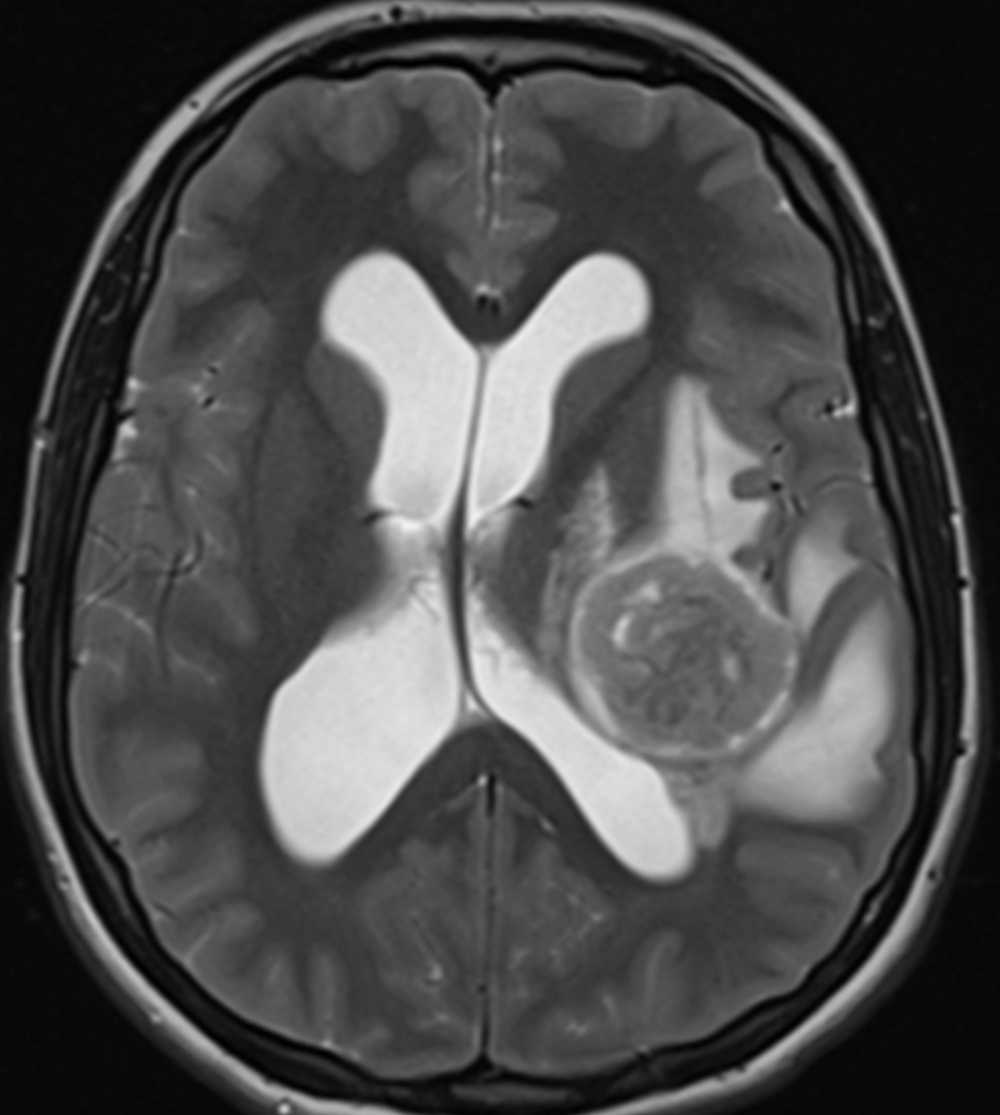
The brain tumour is abnormally malformed tissue that is growing fast (white straight arrow).
The abnormal tissue is compressing over the adjacent tissue.
The tumour causes extra water in the adjacent brain tissue.
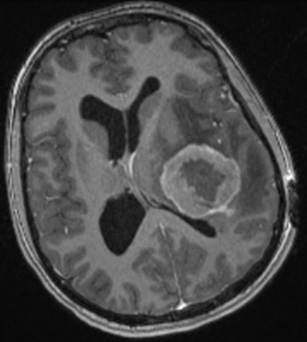
The abnormal extra cells of the tumour need more nutrient which means more vessels are needed. Therefore after injecting dye in the patient’s vessel tumour becomes brighter than adjacent tissue due to more vessels within it (bent arrow).
The interval disc sometimes becomes dry and protrudes backwards into the spinal canal.
the black region is showing the disc compressing over the spinal cord.
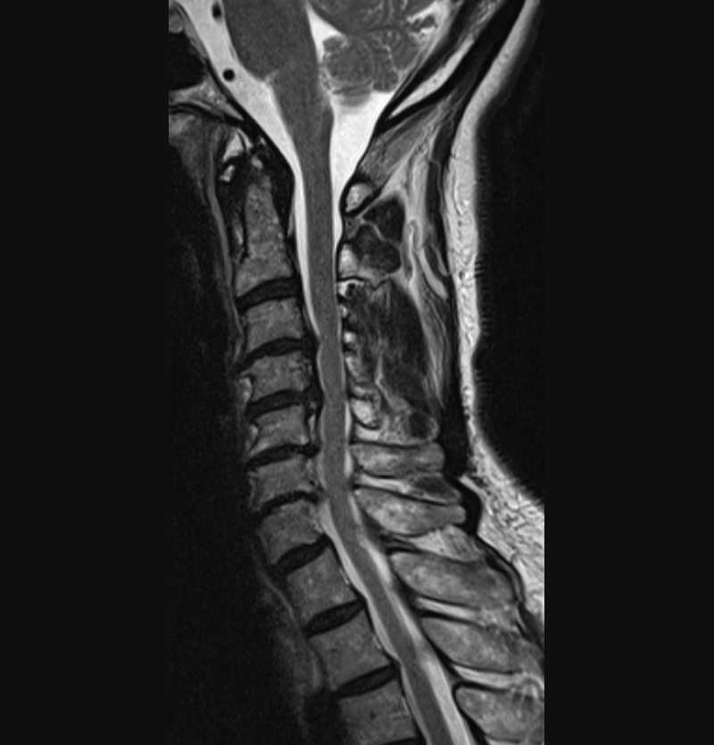
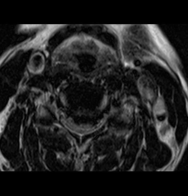
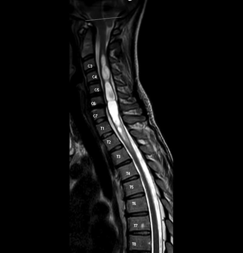
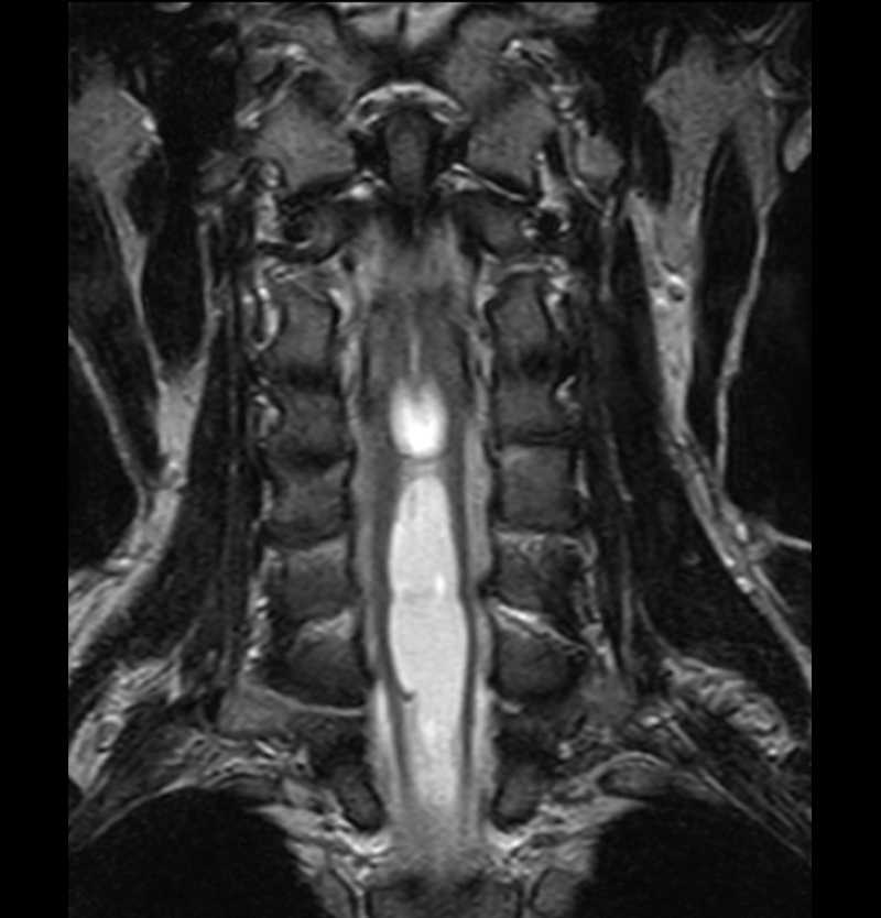
The pointed part of the cerebellum lies below the standard level (red line). This abnormality is called Chiari I malformation and can cause headache and fluid accumulation within the spinal cord known as hydro syringomyelia.
