-
Role of MRI in Headache Evaluation
Headaches are one of the most common healthcare problems and is one of the leading causes for outpatient visits. WHO estimates that three quarters of adults aged 18 – 65 years suffer from headache each year and about 30% reportedly have a migraine episode. While there are multiple treatment options for headaches, the biggest pain point for doctors is the diagnosis. Doctors often use patient’s medical history and symptoms to..Read More -
Role of MRI or CT Scan in Sports Head Injury
Role of MRI or CT Scan in Sports Head Injury A recent research found that sports and related activities contribute to 21% of all traumatic brain injuries sustained by adults in the USA. The only way to ensure quick patient recovery is through fast and accurate diagnosis so that appropriate treatment can be provided. When it comes to head injuries sustained due to sporting activities, there are advanced technologies that..Read More -
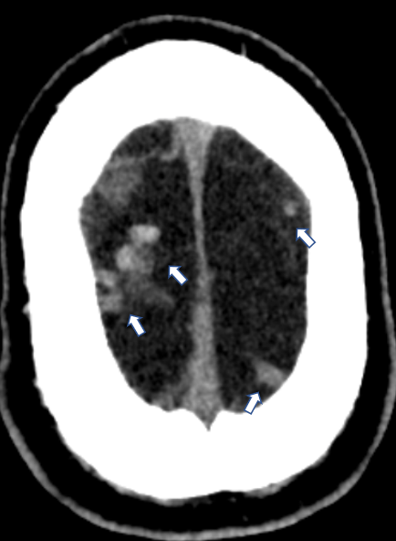
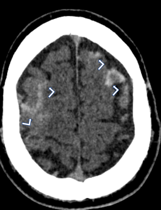
Cortical vein thrombosis
Rarely cortical vein thrombosis (straight arrows) may cause subarachnoid haemorrhage (bent arrows).
-
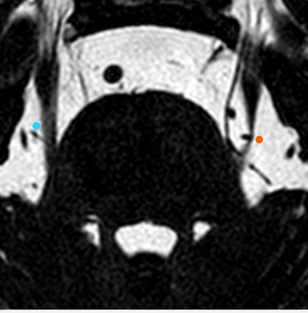
Trigeminal Nerve Neuralgia
The left posterior cerebral artery is indenting the left trigeminal nerve (red dot) causing facial pain. Compare with the blue dot, right normal trigeminal nerve.
-
Does an MRI help in detecting brain tumors?
Magnetic Resonance Imaging, commonly referred to as MRI, is a fairly common test that is used to diagnose a variety of health conditions. Doctors will most likely suggest an MRI when they need to analyze one or more parts of your body including the brain, chest, lungs, and spinal cord among others. Doctors often turn to an MRI test when an X-ray, CT scan or even an ultrasound is unable..Read More
-
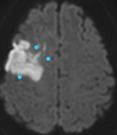
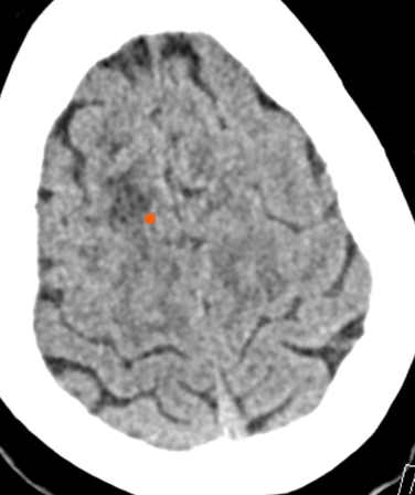
Brain CT Scan v/s MRI Scan
CT: The red dot refers to the dark area of the brain representing acute infarction. MRI: The blue dots refer to the bright area of the brain representing acute infarction. Sometimes the terms CT Scan and MRI Scan are used interchangeably or referred to as simply a ‘scan’. However, there are several significant differences between the two and they are both often used in different situations. I have tried to..Read More
-
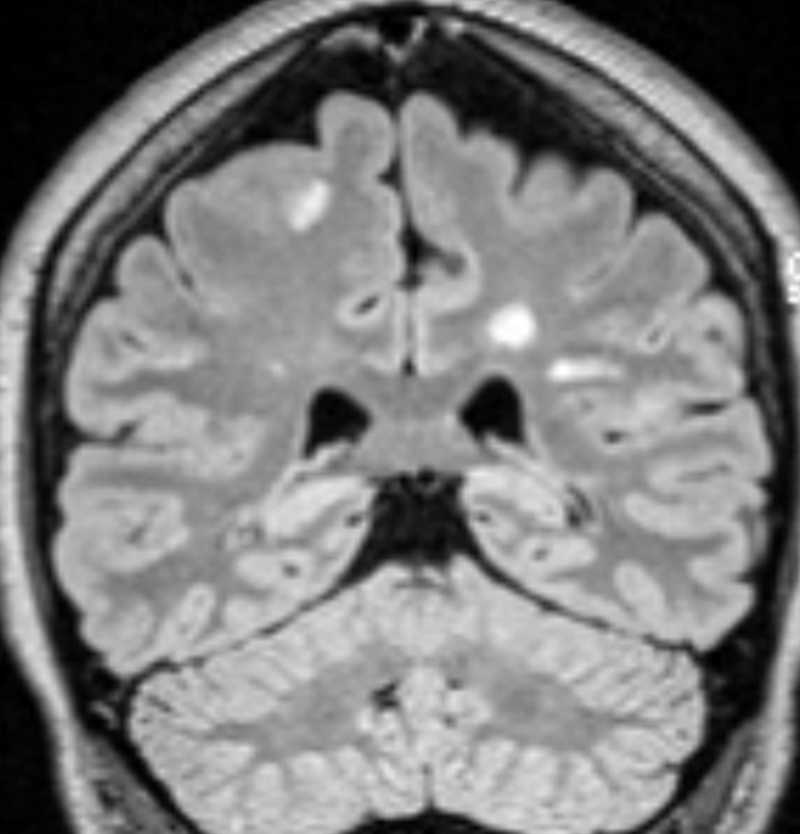
Multiple Sclerosis
The white signal changes within the brain tissue particularly around central brain cavities (ventricles) are showing multiple sclerosis. The number and size of these lesions is usually monitored to decide about choosing the best treatment.
-
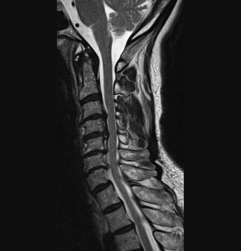
Neck and arm pain
The interval disc sometimes becomes dry and protrudes backwards into the spinal canal. the black region is showing the disc compressing over the spinal cord.
-
Chiari I malformation
The pointed part of the cerebellum lies below the standard level (red line). This abnormality is called Chiari I malformation and can cause headache and fluid accumulation within the spinal cord known as hydro syringomyelia.
-
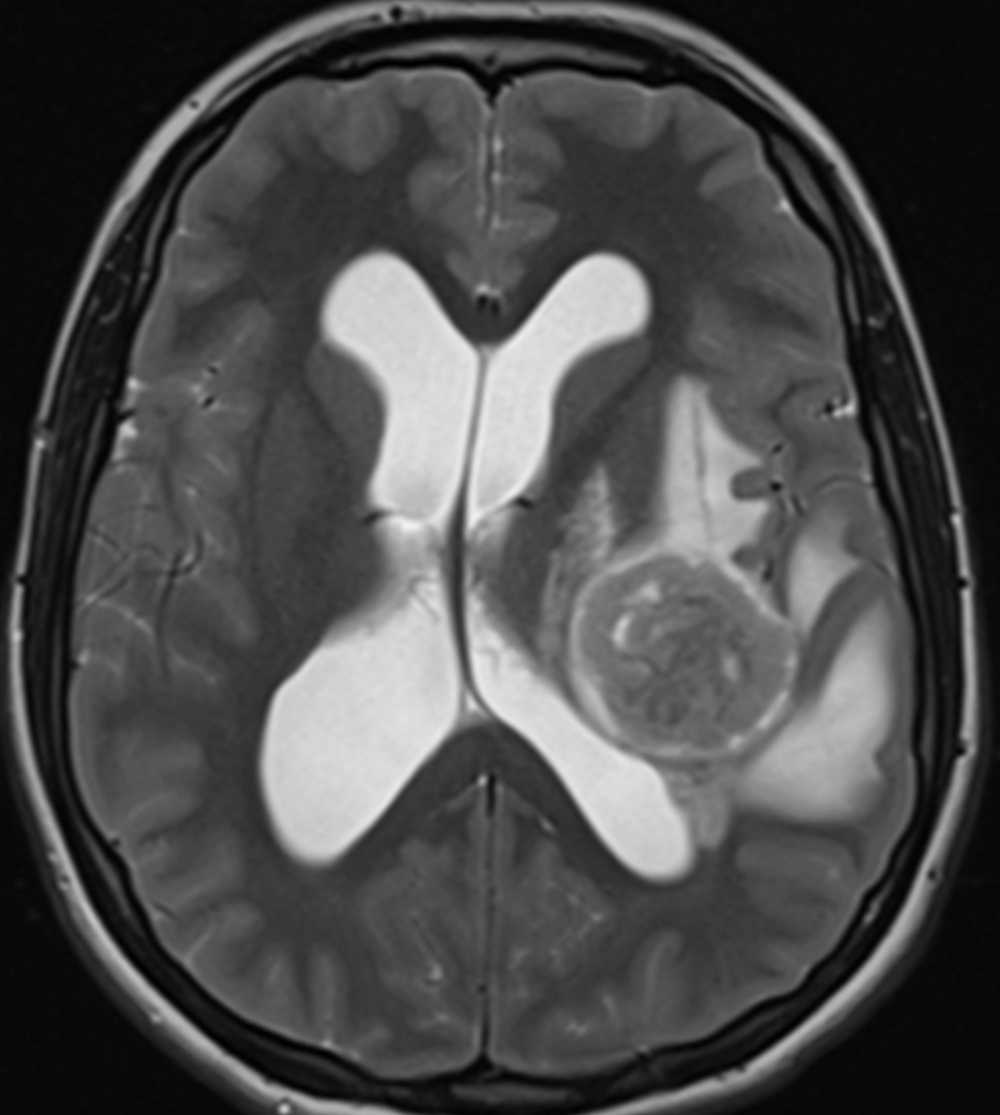
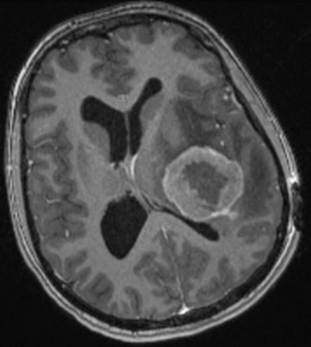
Malignant Brain Tumour
The brain tumour is abnormally malformed tissue that is growing fast (white straight arrow). The abnormal tissue is compressing over the adjacent tissue. The tumour causes extra water in the adjacent brain tissue. The abnormal extra cells of the tumour need more nutrient which means more vessels are needed. Therefore after injecting dye in the patient’s vessel tumour becomes brighter than adjacent tissue due to more vessels within it (bent..Read More